We integrate multiscale 3D imaging, spatial omics, artificial intelligence, high-performance computing, and clinical medicine — building comprehensive brain maps that turn complex biological data into biomedical intelligence.
From molecules to systems, we visualize the brain in three dimensions across multiple spatial scales.
We map genes, proteins, and cells in their native spatial context to reveal the language of the brain.
We develop AI models and scalable pipelines to transform massive imaging and omics data into insight.
We connect brain maps with clinical knowledge to advance precision medicine and brain health.
The Brain Spatiomics Center (BSC) at China Medical University integrates multiscale three-dimensional imaging, spatial omics, artificial intelligence, high-performance computing, and clinical medicine to transform biological structure into digital intelligence.
BSC is designed not simply as an imaging core, but as a discovery and translation platform for understanding how the brain's physical organization gives rise to computation, memory, behavior, and neurological disease.
Spatiomics is the Center's organizing principle. Structural wiring, gene expression, protein localization, electrophysiology, clinical phenotype, and longitudinal disease trajectories are brought into a common spatial framework. This enables biological specimens to become standardized, computable models that can be explored by scientists, interpreted by clinicians, and reused by AI systems.
An integrated research architecture spanning international outreach, data and AI, ethics, imaging factory, functional applications, and clinical translation.
Positioning CMU as a hub for brain spatiomics in Asia and globally — fostering collaboration, exchange, and resource integration.
Developing AI pipelines and high-throughput automation for neuroscience data at petabyte scale.
Integrating philosophy, humanities, and technology for AI medicine, neuroethics, and tech governance.
High-throughput, high-resolution imaging for multiscale observation and connectomics research.
Converting structural atlases into functional understanding — bridging basic science and clinical research.
An integrated clinical, translational, and basic research team to unlock complex neurological conditions.
Reveal how molecules, cells, and circuits generate perception, memory, decisions, and behavior in model animals.
Create AI-enabled, high-resolution maps that connect tissue architecture with diagnosis and treatment.
The Center's operational engine is the AI Bioimaging Foundry — an AI-orchestrated, closed-loop infrastructure that moves from specimen to validated biological knowledge. Standardized sample preparation, large-volume optical imaging, AI super-resolution, Multiview Expansion Tomography (MExT), AI reconstruction, GPU computing, curated knowledge bases, and physician-led validation are integrated into one reproducible pipeline.
MExT supplies high-resolution, near-isotropic three-dimensional reference data; AI/HPC converts these data into aligned volumes, segmented cells and processes, candidate synaptic features, and reconstructed circuits. Standard protocols, quality-control checkpoints, and data provenance make the workflow scalable, comparable, and reusable.
BSC develops a cross-scale framework that connects molecular identity and cellular morphology with circuit architecture, computation, disease, and precision medicine.
Build AI super-resolution atlases of the human brain, beginning with hippocampal and cortical circuits and progressing toward multi-centimeter and ultimately whole-brain mapping.
Learn MoreIntegrate Drosophila, mouse, and human datasets to identify conserved and divergent principles of circuit organization, neural computation, learning, memory, and individualized behavior.
Learn MoreMap cellular vulnerability, circuit disruption, and molecular pathology in epilepsy, neurodegenerative disease, and brain tumors.
Learn MoreCreate AI-ready 3D reference datasets, scalable reconstruction pipelines, and interoperable knowledge platforms for terabyte- to petabyte-scale brain data.
Learn MoreTranslate spatial brain maps into physician-in-the-loop tools for biomarker stratification, treatment planning, and patient-specific digital brain models.
Learn More
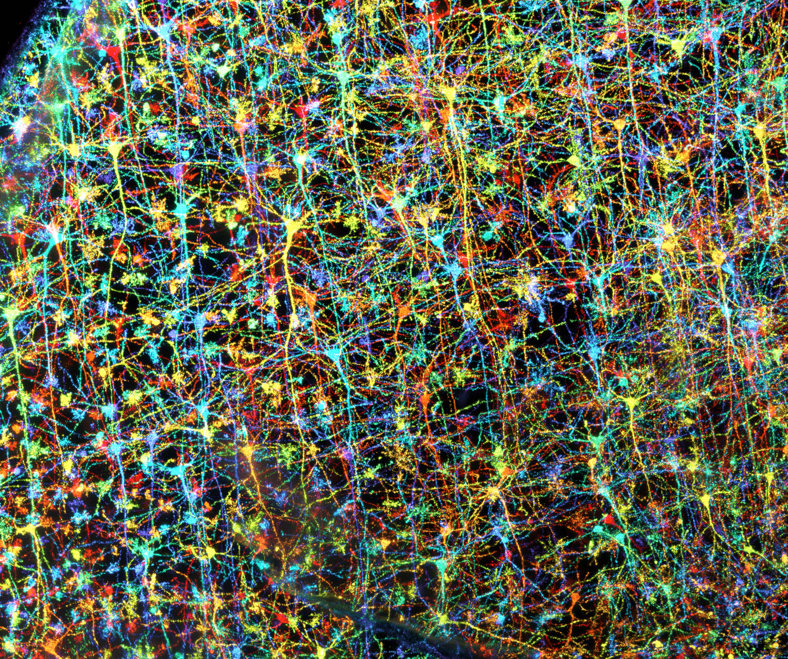
Golgi labeling combined with sevenfold expansion microscopy demonstrates BSC's ability to visualize dense neuronal morphology in clinically relevant human tissue.
Why it matters. The dataset reveals separated neuronal somata and their fine processes within densely labeled cortical tissue (Figure 1), providing morphology-rich reference data for AI segmentation, connectome reconstruction, cross-sample comparison, and human brain atlas development.
This architecture is deliberately bidirectional: high-resolution imaging and molecular measurements provide ground truth for AI, while AI identifies objects, regions, and features, and generates hypotheses that can be returned to imaging, pathology, and clinical experts for validation. The result is an iterative physical–AI workflow rather than a one-way image-analysis pipeline.
Overcomes axial resolution limits in large-volume tissue microscopy, delivering high-throughput and near-isotropic 3D reconstructions across multi-centimeter biological samples.
Leverages high-resolution ultrastructural reference data to guide AI-assisted feature inference across entire tissue blocks, linking micro-architecture to organ-scale contexts.
Converts static histological images into versioned, standardized digital assets — combining aligned volumetric data, automated segmentation, spatial omics, and connectomic graphs.
Extracts principles of memory, sensory processing, and decision-making from experimentally tractable Drosophila circuits to inform human cognitive architecture and neuromorphic AI.
BSC's primary outputs are not images alone, but quantitative and reusable models: connectome maps, molecularly annotated three-dimensional atlases, validated AI models, disease-specific spatial signatures, and computable representations of individual brains.
These resources will reveal vulnerable cell populations, clarify circuit-level mechanisms, identify therapeutic targets, strengthen interpretation of clinical specimens, and provide a structural foundation for precision diagnosis and treatment.
The scale of brain data also creates a frontier for computer science. Reliable analysis of terabyte- and petabyte-scale datasets requires advances in automated acquisition, data movement, distributed inference, uncertainty-aware reconstruction, and human–AI collaboration. In return, decoding the brain's efficient and robust computational architecture may inspire more energy-efficient and trustworthy artificial intelligence.
中國醫藥大學以守護人類健康為使命。在蔡長海董事長的前瞻領導下,我們整合卓越學術、臨床醫療與生醫產業資源,全力打造兼具科研實力、人文關懷與國際影響力的世界級醫學大學。
面對 AI 與生命科學融合的新時代,本校聚焦四大核心領域,推動跨領域創新研究。
以「人才為本、科技驅動、國際接軌、轉譯加值」為策略核心,透過 DNA 引擎建構頂尖跨領域研究平台。
我們積極建構 Smart AI University,推動 AI 深度融入教學、研究與行政。
結合人類創造力與 AI 運算能力,培育兼具專業素養、獨立思考、創新能力與倫理責任的跨域人才。
推廣校園 AI 助理與 AI 虛擬助教,提升整體教學與營運效能。
結合跨域課程、健康照護與在地需求,讓 AI 素養自然融入學習與生活實踐。
展望未來,中國醫藥大學將持續深化國際合作,串聯學研、醫療與產業資源,打造具國際競爭力的生醫創新生態系。我們誠摯邀請全球優秀學者、研究人員與青年人才加入中國醫藥大學——攜手探索生命科學前沿、突破科研疆界,共同開創生醫科研新典範!